Andrew Williams investigates the application of microscopy for disease diagnosis
Andrew Williams investigates the application of microscopy for disease diagnosis.
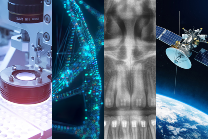
In recent months, a number of innovative projects have demonstrated the utility of cutting-edge microscopy applications for disease diagnosis. So, what approaches and techniques are being adopted? And what technology and equipment do these applications use?
Innovative lensless platform to improve disease diagnosis
The trade-off between the resolution and the imaging field of view is a long-standing problem in traditional microscope systems. To see a small feature with details, an observer must use a high-resolution objective lens with a reduced field of view. However, as Dr Guoan Zheng, United Technologies Associate Professor in the Departments of Biomedical Engineering and Electrical and Computer Engineering at the University of Connecticut (UCONN), explains, this is a ‘major inconvenience’ for life scientists and pathologists who rely on microscopy to analyse and diagnose disease, since prepared tissue samples ‘have dimensions in the centimetre range.’
In an effort to address this trade-off, Zheng and his team have devised an innovative lensless platform dubbed ‘near-field blind ptychographic modulation.’ Rather than using lenses to magnify specimens, the approach instead relies on the placement of a thin diffuser in-between the specimen and the image sensor in a lensless setting. The diffuser is then randomly scanned to different positions while the sensor acquires the images. The captured images during the scanning process contain the encoded complex object information that will later be used to recover a high-resolution, large field of view image of the specimen.
“At the heart of our object recovery process is a microscopy technique called ptychography. Light detectors such as image sensor and photographic plates measure only intensity variations of the light that hits them; in the process of recording, they lose the phase information, which characterises how much the light is delayed through propagation,” says Zheng.
Phase retrieval to recover lost information
Ptychography, originally developed for electron imaging, is a phase retrieval approach that recovers the lost phase information from many distinct intensity measurements. In the Connecticut lensless imaging platform, the sample, the diffuser and the image sensor are brought close to each other. This configuration allows Zheng and his team to use the entire sensor area as the imaging field of view, which is in the region of 50 times larger than that of a regular microscope platform while retaining a similar resolution. As Zheng explains, the word ‘blind’ in ‘near-field blind ptychographic modulation’ has two implications. Firstly, it implies the recovery of both the high-resolution complex object and the diffuser profile at the same time. Secondly, it means no prior information of the positional shift of the diffuser is needed. Instead, the shift of the diffuser can be recovered from the captured raw images via image cross correlation.
“In our implementation, we use a low-cost, do-it-yourself stage to move the diffuser to different x-y positions,” he says.
Another limitation of conventional optical microscopy is the short depth of field of the high-resolution objective lens. For example, Zheng observes that the depth of field of a 20×, 0.4 NA objective is ‘a few microns.’ Acquiring an image with such an objective requires placing the sample exactly at the focal position – otherwise, the captured image will not be able to resolve the fine details of the specimen. In contrast, the lensless imaging platform developed by the Connecticut team allows users to digitally refocus to any axial plane after the data has been acquired.
“The combination of wide field of view, high resolution, and long depth of field of the lensless imaging platform promises substantial gains for digital pathology. This lensless imaging technique may potentially free pathologists from hours bent in front of the microscope, manually focusing the objective lens,” says Zheng.
“Our next step is to work with our colleagues in the UConn Health Center to test its application for digital pathology. We are also testing several different image sensors with smaller pixel sizes to further improve the resolution,” he adds.
Holotomography microscopes and disease diagnoses
Also in the USA, a team of researchers at the University of Illinois at Urbana-Champaign have developed a technique that hopes to ‘bring cancer diagnosis into the digital era’ by pairing infrared measurements with high-resolution optical images and machine learning algorithms to create ‘digital biopsies’ that correlate closely with methods traditionally used in pathology and perform better than cutting-edge infrared microscopes. The hybrid microscope was created by pairing an infrared laser with a specialised microscope lens, known as an interference objective – and adding them both to an optical camera. The results of a digital biopsy of both healthy and cancerous breast tissue samples correlated closely with the results of a traditional one. The research team also found that the infrared-optical hybrid device performed had much larger coverage, as well as greater consistency and higher resolution than modern infrared microscopes.
Elsewhere, the University of Ulsan College of Medicine and Asan Medical Center in South Korea have successfully deployed a Tomocube holotomography microscope to improve the diagnosis of blood cancers. A team of researchers measured the 3D tomogram of individual red blood cells (RBCs) from a patient diagnosed with myelodysplastic syndrome (MDS) and reported that around 30-40% of RBCs in the patient exhibited abnormal cup-like shapes.
As part of the project, a Tomocube HT microscope was used to measure high-resolution, three-dimensional label-free images of individual RBCs. Holotomography (HT) is a laser interferometric technique to measure the 3D tomogram of cells and tissues without labelling or staining. By employing a method broadly analogous to the use of X-rays in CT scans, HT uses a laser beam and reconstructs a non-invasive tomographic visualisation of a sample from the measurements of multiple 2D holographic images. Compared to conventional microscopic methods, HT utilises the refractive index distribution of a biological sample as an intrinsic imaging contrast, thus enabling label-free and quantitative bioimaging.
“For example, in laboratory medicine, complete blood count (CBC) analysis or microscopy imaging of a staining blood smear slide is routinely performed. However, none of these techniques can provide detailed information about tomograms of individual cells … or in-depth assessments of high-resolution 3D images of blood cells, such as with these cuplike shapes,” says YongKeun (Paul) Park, Chief Technology Officer at Tomocube.
Because of these limitations, Park observes it becomes increasingly important to analyse individual RBCs or white blood cells (WBCs) to make ‘earlier and effective diagnoses of several blood-related diseases.’
By enabling such analysis, he also points out that HT possesses a range of potential benefits for clinical applications, including the fact that rapid 3D images can be visualised in ‘tens of seconds’ without time-consuming sample preparations or staining procedures. He also points to the high resolution 3D capabilities of the Tomocube HT model – with a lateral resolution of 0.11 μm and axial resolution 0.36 μm – as well as its quantitative imaging capability, meaning that refractive index values of RBCs can be directly related to clinical Hb parameters such as mean corpuscular haemoglobin (MCH) or mean corpuscular haemoglobin concentration (MCHC).
Moving forward, Park also highlights the fact that the ‘powerful label-free 3D imaging capability’ of HT means its use can be extended for the diagnosis of other diseases, especially when combined with artificial intelligence approaches.
“For example, it has been shown that HT can be potentially used for the diagnosis of infectious diseases or immune-related disorders. Currently, Tomocube collaborates with several medical hospitals and is working on validating the concepts in clinical environments,” he adds.