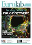
Beamline scientists, Maria Harkiolaki and Ilias Kounatidis, review a new microscopy platform that specialises in imaging the fine details of cellular ultrastructure and chemical localisation in 3D
At the UK’s synchrotron, Diamond Light Source, a novel combination of imaging on beamline B24 is enabling high-resolution 3D imaging of the cellular universe. It uses a partnership of two cutting-edge methods: soft X-ray tomography (SXT) that delivers 3D scans of structures inside and across cells to a resolution of 25nm; and structured illumination microscopy (SIM), which pins chemical information on those scans through fluorescence and super resolution imaging. This combined correlative light microscopy and X-ray tomography (CLXT) platform can deliver content beyond the reach either method can deliver alone.
Within this scheme, correlated 3D imaging data is acquired through an established workflow that sees a sample go from one microscope to the other without any mechanical or chemical processing. This, in turn, ensures that the 3D cellular data captured in one microscope absolutely corresponds to the 3D data captured on the next, making the association of features of interest unambiguous and, therefore, immediately credible as it circumvents the need to infer similarities.
Key to success is sample cryopreservation
Beamline B24 is a Phase III beamline and laboratory that set out to deliver much needed 3D imaging across resolution scales and capture data deep inside cells at near physiological conditions. However, this could only have been achieved through sample cryopreservation. So the journey a sample takes through imaging at beamline B24 begins when it is rapidly plunge frozen in cryogenic liquid. This immobilisation step is required to preserve delicate cellular structures in a state representative of the point in time they were harvested.
Cryogenic temperatures complicate microscope design and sample handling but allow researchers to use intense X-ray and laser light regimes to see features in great detail to a few tens of nanometres even if they reside deep inside cells. For reference, a single Covid-19 particle is just over 100nm in diameter. These features can be delineated in X-ray projections and, if they are tagged with fluorophores that relate information on their current state, they can be chemically characterised. The development of this correlative platform has only recently been fully documented in a publication[1], using as a proof-of-concept this experimental approach to study viral infection processes in human bone osteosarcoma cells. In it, the B24 CLXT shed light on reovirus behaviour during the early stages of infection and provided data on the mechanism that it employs to travel and propagate within human cells.
The B24 team are experts in the preparation, handling and transfer of cryopreserved samples. They work closely with the user community to help researchers get the best results possible and plan experiments that make the best use of the available technology.
Instrumentation and applications of beamline b24
The beamline delivers SXT using radiation from a bending magnet at the synchrotron, which is then reflected and conditioned via a number of specially designed mirrors and ultimately it is delivered as a highly focused 500 eV beam to an X-ray microscope (UltraXRM-S220c, Zeiss) at 500eV. Samples are mounted on round 3mm wafers and kept at cryogenic temperatures at all times. Data collection can be driven in person or remotely and raw imaging data enters an automated pipeline for reconstruction and processing.
For SIM experiments at cryogenic temperatures, the cryoSIM microscope was developed through the collaborative work of the B24 team and Micron, the microscopy facility at the Biochemistry department of the University of Oxford. Bringing together the resident expertise on cryo-handling, sample optimisation and data collection and the wealth of knowledge from microscopy developers in academia resulted in the construction of a user-friendly and accessible super resolution fluorescence module that perfectly complements the SXT facility. It is always used before exposure to X-rays depletes fluorophore capabilities and provides 3D data on chemical localisation based on the fluorescence of the trackers used. This data can show details even below 200nm resolution and the reconstruction process delivers high-contrast, low-background imaging beyond the diffraction limit of the light used.
The technology at B24 has been used so far to study eukaryotic cells, algae, archaea, pathogens (parasites, viruses and bacteria) and their interactions with host cells, stem cell development and differentiation, autoimmune pathologies, biomaterials and their biological applications as well as anticancer drug and vaccine effects in cells. Because of the way samples are cryopreserved for CLXT, intracellular processes can be track-and-traced in 3D and at high resolution without disturbing delicate cellular structures, which allows the capture of dynamic processes such as drug delivery, pathogen clearance and cell signalling.
The power of CLXT has been recently demonstrated in a collaboration with the Kennedy Institute of Rheumatology in Oxford University[2] where, combining a series of imaging techniques (confocal, dSTORM and X-ray microscopy), cytotoxic T lymphocytes (CTLs) were shown to generate and transfer distinct cytotoxic multiprotein complexes, called supramolecular attack particles, to eliminate target cells. According to Professor of Immunology at the Kennedy Institute, Michael Dustin, “Beamline B24 is a very exciting resource to study exchange of information in the immune system. Matching the 25nm resolution of SXT with similar resolution of specific molecular assemblies by super resolution fluorescence such as SIM and STORM, will open up many fundamental questions in immunology and cell biology.”
A number of industrial groups have also used the beamline to evaluate processes and materials. Recent work involved the evaluation and application of new vaccine formulations in mammalian cell lines to confirm antigenic potential and overall effect on host cell physiology (manuscript in preparation). According to Dr Claire Pizzey, deputy head of the industrial liaison group at Diamond: “The scientific insight gained using the B24 correlative microscopy approach has been of great value in the areas of vaccine development and novel therapeutics, feeding into our clients’ R&D pipelines in a timely manner. The full-service approach, where we support our clients through experimental design, data collection, analysis and reporting, is of particular interest to our clients who recognise the benefits of the technique but many have no prior experience.”
Accessibility of correlative imaging for academia and industry
Continued demand to deliver correlative imaging at the cellular level in complex biological systems is currently driving further developments at the beamline. This will ensure high-quality data is accumulated faster and more reliably and it is anticipated that many otherwise challenging projects will benefit from its application in the future. The microscopes at beamline B24 are accessible to both academia and industry via well-defined access routes. For academic groups, access requires the submission of a proposal: a ‘rapid access’ one for proof-of-principle studies or a ‘standard’ one for established projects; the latter enters a competitive access process that involves peer-review and award of usage shifts. Industrial groups on the other hand access the beamline via the industrial liaisons team at Diamond. It is recommended that potential users contact the B24 team to discuss requirements and plan their experiments in way that takes full advantage of what is on offer. During the Covid-19 pandemic users are still able to send their samples to B24 and conduct data collection remotely while all the necessary protocols and software instructions are fully accessible online.
Beamline b24 is enhancing our understanding of the cellular world
The correlative microscopy platform available at B24 is a new tool that has been developed specifically to promote and enhance our understanding of the cellular world and its potential. The technology on site is user-friendly and accessible and remains dynamic as it is upgraded constantly in response to the latest demands of the wider biomedical community.
References[1] Kounatidis I, Stanifer ML, Phillips MA, et al. 3D Correlative Cryo-Structured Illumination Fluorescence and Soft X-ray Microscopy Elucidates Reovirus Intracellular Release Pathway. Cell. 2020;182(2):515-530.e17.
[2] Bálint, S et al. Supramolecular attack particles are autonomous killing entities released from cytotoxic T cells. Science(New York, N.Y.) vol. 368,6493 (2020): 897-901.
Maria Harkiolaki is principal beamline scientist, B24 and Ilias Kounatidis is beamline scientist, B24, at Diamond


