The microscope can image entire 3D datasets at a precise moment in time
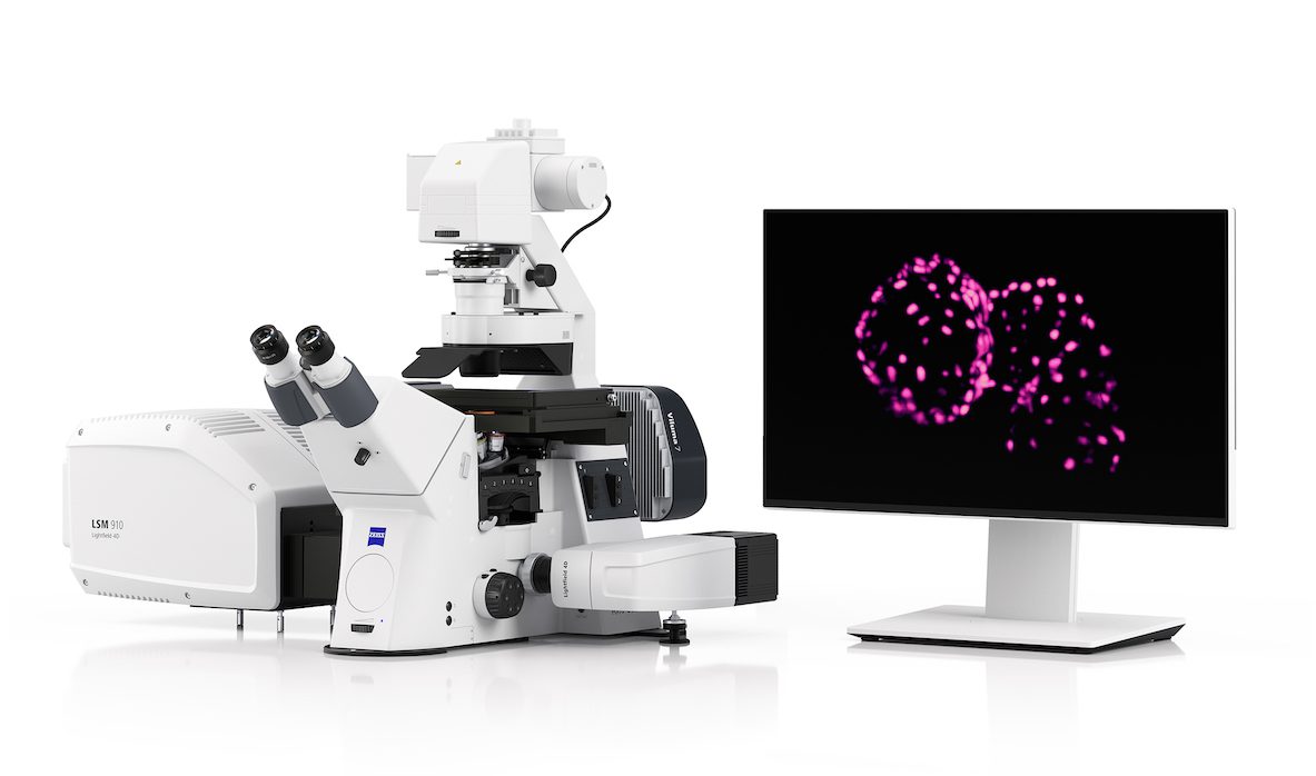
Optical systems developer Zeiss has recently launched Lightfield 4D, a new microscopy technology based on the light-field principle.
“This innovative system enables researchers to capture biological processes in real-time, providing unprecedented insights into the dynamic nature of life,” said Zeiss head of life sciences microscopy Dr Bernhard Zimmermann.
Lightfield 4D images entire 3D datasets at a precise moment in time, combining volume and time to explore biological processes. The instant volumetric high-speed images can capture dynamic processes of life in complete samples with high temporal resolution.
The technology enables researchers to more accurately observe living organisms and biological events such as physiological and neuronal processes. Lightfield 4D has applications in neuroscience, cancer research and developmental biology.
Traditional optical sectioning techniques rely on sequential image acquisition to create Z-stack images. Sequential image acquisition introduces time delays, compromising the spatiotemporal accuracy of data.
Using a micro lense array, Lightfield 4D facilitates the capture of multicolour volumes of large samples. It generates 37 individual images simultaneously to capture 3D information in real time. This allows of the creation of Z-stacks through advanced deconvolution-based processing, achieving up to 80 volume Z-stacks per second.
Since a complete Z-stack is captured in a single illumination event, light exposure and phototoxic effects are minimised, allowing for long-term imaging of entire living organisms at high temporal density.
Lightfield 4D is integrated as an imaging mode into the Zeiss LSM 910 and LSM 990 confocal microscope systems.
Late last year, Zeiss introduced the Crossbeam 550 Samplefab, a focussed ion beam scanning electron microscope.