Lighting the way: using fluorescent lipid nanoparticles to identify promising formulations for oncology treatment
Messenger ribonucleic acid (mRNA) technology became widely known following its widespread rollout in vaccines during the COVID-19 pandemic. However, mRNA also has potential uses in oncological treatments. Early results are promising, but there is work to be done to demonstrate full clinical effectiveness, especially regarding cancer cell specific targeting.
Tebubio offers contract research services in RNA-based therapeutic discovery and has leveraged its platforms to develop fluorescent lipid nanoparticles (LNPs) that are designed to facilitate image-based tracking of RNA delivery. This has enabled identification of the most suitable formulations for LNP-based mRNA delivery in 3D tumour spheroids, paving the way for easy monitoring of LNP delivery, and for future personalised RNA-based oncological treatments.
Despite the fact that LNPs are one of the most successful nano-delivery vehicles for RNA therapeutics,ii LNP delivery of RNAs is not without its challenges. Studies about RNA delivery via LNP suggest that as little as 2% of RNA particles are released into the cytoplasm of the cell.iii This process, known as endocytosis, is essential for the RNA to facilitate desirable protein synthesis within the ribosomes. Therefore, LNP formulation requires careful consideration to ensure this process takes place efficiently. By incorporating a fluorescent dye into an LNP, transfection and RNA expression across different LNP formulations can be monitored.
This project aimed to identify the best formulations to target specific cancer cells by transfecting HCT116 cells (a colorectal cancer cell line) with mRNA encapsulated in different LNPs. This requires a 3-step process – RNA production, RNA encapsulation in LNP, and transfection of encapsulated RNA in a colorectal cancer in vitro model. This enabled identification of the best delivery formulation to express the mRNA in its targeted cells.
From RNA production to 3D in vitro modelling
To identify the most promising LNP formulations for mRNA-based therapeutics, the project aimed to assess the best formulation recipe which would allow high levels of transfection of an mRNA coding for the eGFP protein. To do so, eGFP mRNA was produced by in vitro transcription (Fig. 1, left). Then, the eGFP mRNA was encapsulated into seven formulations of LNPs, with each formulation differing by ionisable and structural lipids (Fig. 1, centre). To help monitor transfection success, a fluorescent dye (Cy3) was added to a structural lipid within all the LNP formulations.
LNP formulations containing eGFP were first transfected in HCT116 2D cell cultures. These were then screened to see which LNP formulation showed high transfection efficiency and expression of eGFP in the 2D cultures, to determine the most promising formulation for 3D in vitro modelling (Fig. 1, right).
Once the most successful mRNA-based LNP was tested in 3D in vitro models, the fluorescent dye continued to facilitate monitoring of LNP transfection and eGFP expression. This testing identified the most promising formulation for further in vitro and potentially in vivo studies (Fig. 1, right).

Figure 1: Study workflow from RNA production to readout after incubation on 2D or 3D culture of HCT116 cells, including formulation in LNP and quality controls.
In vitro modelling
Once the seven LNP formulations were established, all seven formulations were screened to assess their ability to deliver the eGFP mRNA into the 2D HCT116 cells. Transfection was performed using 200 ng of the eGFP mRNA, and eGFP expression was assessed 24- and 48-hours post transfection (Fig. 2).
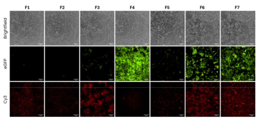
Figure 2: Transfection efficiency of 7 different LNP formulations in HCTT116 culture models after 48 hours. Cells were transfected with 200 ng of eGFP-mRNA encapsulated in LNPs. Acquisitions were performed to monitor protein expression (eGFP), cell survival (Brightfield) and LNP distribution (Cy3).
The results clearly show not only which LNP formulations resulted in successful transfection, but also which formulations were the most effective in facilitating eGFP expression. Figure 1 shows that formulations (F) 4,6, and 7 were the most effective in both transfecting the cells and expressing the eGFP, therefore presenting as the most promising formulations for 3D testing.
As one of the most promising formulations in the 2D model, the F4 LNP formulation was chosen to test whether this formulation facilitated the transfection and expression of eGFP in a 3D in vitro model. This experiment aimed to prove that LNP delivery of mRNA could be a potent method for targeting colorectal cancer models.
Hence, eGFP mRNA was produced and encapsulated into the F4 LNP at different concentrations. Then, 3D tumour spheroids of HCT116 were transfected with the encapsulated eGFP mRNA, and the eGFP and cell death levels (using Cytotox) were monitored for three days (Fig. 3a).
Figure 3: (a) Representative images of HCT116 spheroids transfected with different concentrations of

Figure 3: (a) Representative images of HCT116 spheroids transfected with different concentrations of eGFP (green) encapsulated in F4 LNP at 72h post transfection. Cytotox (red) was used to assess apoptosis levels in the spheroids. (b) Levels of eGFP were quantified and reported in a graph. n=2 wells with between 20 to 24 spheroids. Scale bar = 500 μm.
The results showed that the eGFP was successfully expressed in a concentration dependent manner (Fig. 3b) in the tumour spheroids via the F4 LNP without inducing significant cell death as compared to control (no transfection, Fig. 3a). This demonstrates that mRNAs can be successfully encapsulated, delivered and expressed into cancer cells using LNP for encapsulation – a promising result for the future of colorectal cancer treatment tools.
Next steps for mRNA in personalised oncology treatments
The use of fluorescent lipids verified the successful delivery of mRNA encapsulated in specific LNPs as a potential treatment for colorectal cancer. This represents a breakthrough in the future of personalised oncology treatment, as this technology can be applied to patient-derived tumour spheroids, to identify personalised formulation recipes to target specific cancer cells in patients.
References i Nogrady B. mRNA technology helps reinvigorate the hunt for cancer vaccines. Nature. 2025 Apr;640(8060):S54-S56. doi: 10.1038/d41586-025-01151-7. PMID: 40269290. ii Jung HN, Lee SY, Lee S, Youn H, Im HJ. Lipid nanoparticles for delivery of RNA therapeutics: Current status and the role of in vivo imaging. Theranostics. 2022 Oct 24;12(17):7509-7531. https://doi.org/10.7150/thno.77259. PMID: 36438494; PMCID: PMC9691360. iii S. Chatterjee, E. Kon, P. Sharma, & D. Peer, Endosomal escape: A bottleneck for LNP-mediated therapeutics, Proc. Natl. Acad. Sci. U.S.A. 121 (11) e2307800120, https://doi.org/10.1073/pnas.2307800120 (2024).