Raman spectroscopy can provide a faster, non-destructive complement to traditional methods of detection
Early detection is critical to cancer survival, particularly for cervical cancer, the fourth most common cause of cancer death in women around the world. Despite it being largely preventable, the disease continues to claim lives due to the limitations of existing screening methods.
Early warning signs can be found through routine Pap smears and HPV tests, but both have shortcomings. Pap smears rely on visual interpretation, meaning biochemical abnormalities invisible to the human eye often go missing. HPV testing is highly sensitive but lacks specificity, so a positive result could indicate a harmless transient infection as opposed to an actual cancer risk.
Professor Fiona Lyng, manager of the Radiation and Environmental Science Centre at Technological University Dublin, has been on a mission to improve detection through optical spectroscopy. Over two decades, her team has pioneered Raman spectroscopy for early, objective detection of precancerous changes in cervical cells, providing a faster, non-destructive complement to traditional methods.
Raman spectroscopy
Lyng said, “Early detection is key for any cancer. If you can pick up precancer, you can intervene before it spreads.”
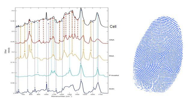
Raman spectroscopy utilises laser light to measure subtle molecular vibrations in cells, generating a unique biochemical fingerprint, enabling researchers to detect cellular changes before they become visible under a microscope.
Lyng first encountered Rama spectroscopy in 2000, after her lab acquired Ireland's only HORIBA Raman microscope. Through collaborating with clinicians at Dublin's National Maternity Hospital, Lyng's team began analysing cervical tissue samples and saw the technique's diagnostic potential.
The team developed a minimally invasive process compatible with existing screening workflows by adapting Raman spectroscopy for clinical cytology samples.
Clinical-ready protocol
Lyng said, “Developing the protocol took a lot of trial and error. We wanted it to work on standard glass slides so it could be used in clinical labs without changing the workflow.”
Using glass required a 532nm green laser to reduce background fluorescence; this wavelength also introduced interference from haemoglobin. The team was able to overcome this through careful optimisation, resulting in a simple, non-destructive Raman test that complements existing cytology and biomarker analyses.
Clinical studies
In 2022, the group validated their technique in collaboration with the Coombe Women and Infants University Hospital, analysing 662 patient samples. The study, which is the largest of its kind for Raman microscopy in cervical precancer detection, achieved 91.3% classification accuracy between healthy and precancerous samples.
Lyng said, “This accuracy demonstrates Raman’s potential as a powerful diagnostic adjunct."
A partner in discovery
Much of this work is supported by HORIBA Raman instruments, with the Technological University Dublin operating two LabRAM HR800 systems and two HORIBA XploRA Raman microscopes, which have been essential throughout the project.
Lyng said, “The XploRA systems have been incredible. They’re compact, easy to operate, and ideal for hospital labs. Even after 15 years, they still produce high-quality data every day.”
The HORIBA XploRA is a benchtop confocal Raman microscope that combines spectral resolution with user-friendly automation, featuring a modular design that supports a variety of laser wavelengths for flexible, high-sensitivity measurements across life-science and materials applications.
Expanding the frontiers of screening
Lyng's team explored Raman spectroscopy to enhance HPV triage, distinguishing between transient and transforming infections. The team's pilot study showed 85% sensitivity and 92% specificity, suggesting Raman could reduce unnecessary procedures and ensure timely care for high-risk cases.
Beyond cervical cancer, Lyng's team are extending Raman spectroscopy to oral precancer, with collaborations with Dublin University Hospital showing that Raman can effectively differentiate between stages of oral dysplasia. A new Science Foundation Ireland project is aiming to develop saliva-based Raman analysis for non-invasive screening of potentially malignant oral lesions.
Lyng said, “For all these studies, our HORIBA XploRA systems remain essential. They’ve taken us from discovery to real-world validation.”
The future of cancer diagnostics
Lyng's research highlights how light-based technologies can transform cancer detection. By identifying the biochemical signatures of disease before any physical changes occur, Lyng's work offers hope for earlier, more accurate, and less invasive diagnostics.
Lyng said, “As scientists, our goal is simple. If we can detect cancer before it begins, we can save lives. That’s what drives us forward.”
For further information, visit: Detecting early signs of cancer with Raman microscopy – HORIBA