Lung-on-a-chip grows human tissue to see how SAR-CoV-2 cells spread
Despite attempts to maintain objectivity and accuracy, clinical trials are notoriously flawed since traditional in-vitro assays don’t recognise the intricacies of a living being while in-vivo animal models are not 'human' enough to convey the complexity of a living person.
Ultimately, most clinical drug trials fail, reflecting the fact that preclinical tests do not accurately match human results. How can researchers tame some of these variables and produce quick, cost-effective results that advance disease control?
One method is the use of “organ-on-a-chip” (OOC) technology, which grows primary human cells together in specific combinations to replicate human physiology and function in-vitro.
How does OOC technology work?
This OOC model replaces an older method in which cells were cultured under static conditions on 2D substrates coated with either serum or extracellular matrix (ECM) molecules.
These older models were good for observing cells but failed to promote tissue specific functions. The functions among human organs rely on intricate interactions between various specialised cell types at specific interfaces, arranged in complex geometries and operating in specific microenvironments.
According to a whitepaper from a company operating in the field called Fluigent, recent advances in microfabrication, bioengineering and stem cell biology have meant scientists can create OOCs, a 3D cell culture model that replicates these parameters.
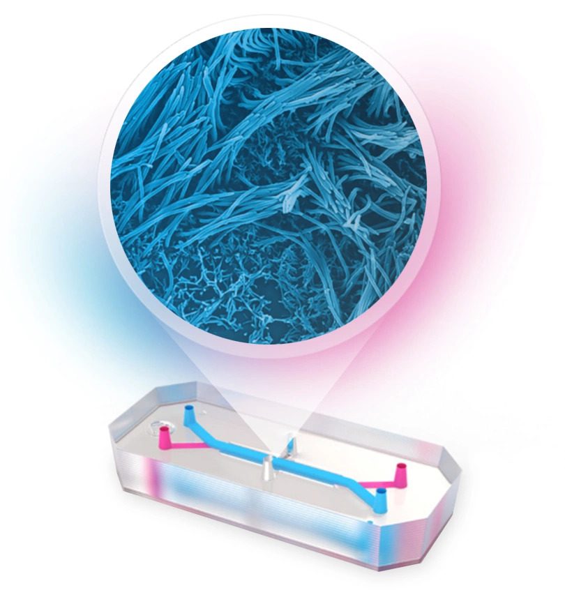
Rather than being miniature organs on a silicon chip soldered to a circuit board, the models are bioengineered devices that use microchip technology design principles. OOCs reproduce key functional aspects of an organ or a tissue, containing structural complexity and incorporating a dynamic fluid flow as well as mechanical stimulations.
OOC's recreate key aspects of human physiology such as the cell-cell interface between an epithelium and the endothelium of organs, the tissue-level organisation of an organ, and the systematic interaction of multiple organs.
Not only does this method help accurately recreate human organ and study tissue components, but it can demonstrate how cells behave over time. OOC technology is particularly useful for studying cancer and inflammatory conditions such as Alzheimer’s and inflammatory bowel disease, because it is now possible to model intricate immune responses. Essentially, the cellular diversity usually seen in-vivo can now be achieved on a small chip.
A new product from the Quadram Institute
In fact, researchers from the Quadram Institute have worked with UK Health Security Agency (UKHSA) to develop a human 'lung-on-a-chip' model. This model recreates how SARS-CoV-2, the seventh known coronavirus to infect people, infiltrates lung cells.
“UKHSA is proud to be supporting this area of research. Systems such as these are becoming increasingly important in understanding the effectiveness of new drugs and play an important role in our preparation for future pandemics,” said Dr Kevin Bewley.
How the lung-on-a-chip works
This model cultivates the cells that line the lungs in humans to be grown in virtually identical conditions, including a stretching motion to mimic breathing and encourage cell development. The cells are grown half in liquid and half exposed to the air, where the virus potentially enters. Upon the virus’s entrance, the model accurately mimics infection, early immune response and the release of new virus particles.
For example, the virus infects the upper airways, but in severe cases, the infection spreads to the lower airways the alveoli, which are small, balloon-like structures that inflate within the lungs when air is inhaled. If the alveoli are damaged, oxygen cannot pass through specialised cells in exchange for carbon dioxide, resulting in respiratory failure.
Professor Nathalie Juge and Dr Tanja Šuligoj worked with biotechnology company Emulate to produce the alveolus lung chip to account for this. The alveolus lung chip was made from human alveoli lining cell and lung microvascular endothelial cells, mimicking alveoli inflation and deflation during breathing.
“We were pleased to be able to pivot our expertise in organ-on-chip models to contribute to the international effort to fight SARS-CoV-2 during the COVID-19 pandemic,” said Juge.
The team worked with UKHSA’s Dr Simon Funnell and colleagues to study the system and ensure its accuracy, finding evidence that the SARS-CoV-2 cells had replicated in all chips tested. The model even reflected the timespan humans often experience, with a high level of the virus found during the first three days.
“The capacity to successfully infect lung-on-chips in Quadram’s CL3 was a milestone and we are pleased to have helped transfer this expertise to UKHSA. This infection model recapitulates clinically relevant effects that can be used to combat emerging infectious respiratory diseases,” added Juge.
UKHSA is continuing to advance this research, analysing how coronavirus can cause more severe disease and how new treatments might work. With this new method, researchers are beginning to understand how SARS-CoV-2 invades the lungs, and how this knowledge could accelerate the drug development process.
With the complexities of the human body no longer off limits, it is possible this research will continue developing and help prepare against future pandemics.