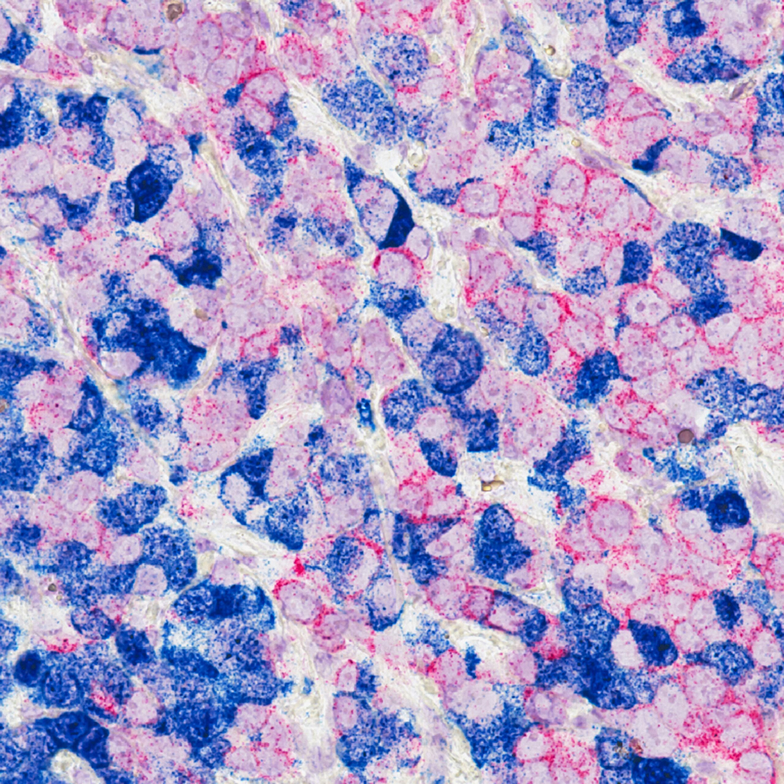
The Atlas H&E-TME provides analyses at single cell resolution in hours
Following the early access pilots with leading biopharma partners, Aignostics has made Atlas H&E-TME generally available, an application for analysing the tumour microenvironment (TME) in H&E images
Atlas H&E-TIME addresses a bottleneck in cancer research by extracting insights from routine H&E slides without specialised staining or multiplexing, and by analysing whole slide images in hours, enabling rapid characterisation of immune cell populations, spatial relationships, and tissue architecture across various cancer types.
Atlas H&E-TME was designed utilising Atlas, the leading foundation model co-developed by Aignostics, Mayo Clinic, and Charité Berlin. It provides detailed readouts for quality control, seven tissue types, and nine cell classes, as well as more than 5,000 higher-order quantitative metrics per image.
The application has been extensively validated across primary and metastatic sites from multiple labs and scanners, ensuring generalisability to real-world conditions. If required, validation metrics can be provided upon request.
Frederick Klauschen, co-founder of Aignostics and director of the Institute of Pathology at Ludwig-Maximilians-Universität München, said, "This is the first time I have seen a model that demonstrates consistently high accuracy across multiple cancer indications with exceptional robustness."
Viktor Matyas, co-founder and CEO of Aignostics, said, "Understanding the TME is essential for developing effective cancer therapies, but traditional approaches are costly, time-consuming, and often limited in scale. Atlas H&E-TME changes this paradigm. Our pilots with biopharma have confirmed we can deliver high-quality results with unparalleled accuracy, speed, and scale, helping researchers get maximum value from every H&E image."