Exploring techniques that provide detailed information on sample topography
The most common modality used in scanning electron microscopy (SEM) is secondary electron (SE) imaging, which detects lower energy electrons emitted from the sample surface owing to interactions with the SEM electron beam. SE imaging provides information about sample topography.
Backscattered electron (BSE) imaging, on the other hand, detects higher energy electrons scattered back from deeper within the sample. BSE imaging reveals sample composition by atomic number contrast with heavier elements appearing brighter.
BEX imaging combines BSE sensors with X-ray sensors placed below the objective lens. The X-ray detectors collect characteristic X-ray emissions that are generated by the sample when irradiated by the SEM electron beam. The X-ray signal is processed to identify and assign colours to the detected elements, which are then combined with the BSE signal to create a final image. BEX imaging provides more information about sample composition and elemental distribution in the same acquisition time as SE or BSE imaging.
Compared with an Energy Dispersive Spectroscopy (EDS) detector, a BEX imaging system offers a higher solid angle, enabling X-ray information collection at normal imaging speeds. The position of the X-ray sensors near the objective lens eliminates shadowing effects and allows for sample investigation at various working distances.
The Unity detector, recognised as one of the top microscopy innovations of 2024 by the Microscopy Today Innovation Awards, combines two types of sensors within a single detector head. It integrates backscattered electron (BSE) sensors and X-ray sensors to provide comprehensive imaging with atomic number and elemental data. The detector is designed for daily imaging, offering flexibility in working distance, the ability to capture data from challenging sample topography, a wide field of view, and compatibility with variable pressure mode for non-conductive samples. The Unity detector delivers reliable and instant imaging results supported by advanced technology and software integration.
Volume Electron Microscopy (vEM): how BEX imaging can accelerate vEM imaging
The Volume EM Technology Forum is a great opportunity for researchers and scientists to learn about the latest advances in volume electron microscopy (vEM). This year, the conference featured presentations from leading experts in the field, as well as technical presentations, posters, panel discussions, and hardware and software demos. Attendees also had the opportunity to network with other researchers and scientists, and to learn about new products and services from exhibitors.
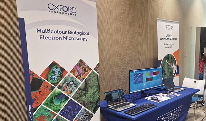
In this year’s event (21st October 2024 – 24th October 2024), the Oxford Instruments team presented a demo of our BEX detector, Unity, where we connected remotely to the Oxford Instruments Innovation Centre, a state-of-the-art facility at the High Wycombe site. We showcased the advantages of BEX imaging for biological and non-biological samples, especially for vEM applications, where the speed and flexibility of BEX for different specimens is key (Figure 1).
With Unity Cartography mode, Oxford Instruments achieved high-resolution chemical mapping of entire biological samples. This workflow would previously have taken several hours to complete (even overnight) and can now be achieved within minutes with BEX imaging, enabling higher throughput and less beam damage for sensitive samples such as cells and tissues (Figure 2).
BEX cartography of cells and tissues
With BEX, new possibilities for vEM are open, for collecting elemental and ultrastructural information simultaneously, without added imaging time. In the example in Figure 3, BEX cartography was used to achieve high resolution images and fast mapping of array tomography brain slices. Large areas were imaged with a relatively short beam dwell time (10µs), which reduces beam damage, drift and resin charging. Elemental information was acquired simultaneously and provided chemical differentiation that can be used to further distinguish between sample features, opening the possibility of improving subsequent segmentation of data. BEX also provided information about strain distribution, which was quantified by the EDS detector. Being able to measure the amount of stain taken up by the sample improves the team’s ability to make direct comparisons between samples enabling them to optimise sample preparation techniques.
For more information visit: http://nano.oxinst.com