Free magazine subscription
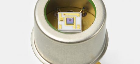
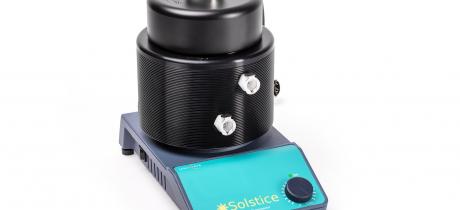
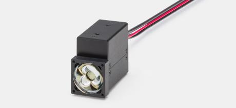
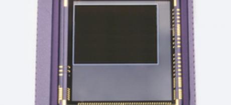
SPAD device will help with low light levels
Available with two photosensitivity settings